IS SYSTEMATIC DETECTION OF VENOUS REFLUX MANDATORY IN
PRACTICE?
Ugur Bengisun, MD Professor of General Surgery
İbn-i Sina Hospital of Ankara University, Dept of General Surgery, Division of Peripheral Vascular Surgery
Ankara/Turkey
Address: Private Office : 1071 plaza 1443 cad No:25A Daire 84 Çukurambar 06510 Ankara Turkey
Telephone: +90 312 466 1656 & 05058555101
e-mails: info@ugurbengisun.com & ugurbengisun@yahoo.com
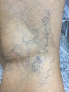
Chronic venous disease (CVD) of lower extremities is a major socio-economic burden with high health care expenditures in many countries. The spectrum of clinical manifestations of CVD ranges from telengiectasias to varicose veins (vv) and severe venous skin changes ie lipodermatosclerosis and venous ulcer. Epidemiological studies have demonstrated that the prevalence of vv as the most common clinical form CVD is around 25-33% in women and 10-20 % in men.
Although the pathogenesis of CVD is not fully or clearly understood, sustained venous hypertension is the result of symptoms or signs of vv which are due to valvular incompetence and venous obstruction. The presence of reflux alone is 80% in patients with CVD while obstruction alone is detected in only 2%. The combination of reflux and obstruction is seen in about 17%. Because of primary valvular incompetence or postthrombotic (PT) damage superficial veins are affected in 90% of lower extremities with
CVD, the deep veins in only 30% and perforating veins in 20%.
In contrast to wide variety lower limb symptoms including aching, heaviness, pruritis, swelling and restless legs many patients with CVD are asymptomatic. Additionally many studies have shown that such symptoms are present in about half the adult population and increase significantly with age. Symptoms of venous disease alone are usually not characteristic. There is no good correlation across clinical symptoms, vv patterns and severity of venous reflux. ¹ Moreover patients with venous aching or swelling cannot be associated with apparent vv during examination. Interestingly similar patients who have minimal or no varicosities can show major venous reflux by duplex scanning. Therefore symptomatic patients should be assessed with great attention starting from history taking and physical examination points in search of characteristic features of alternate ischemic, arthritic or neuralgic causes for pain. This will lead to appropriate diagnostic investigations and treatment modalities.
The agreement between symptoms and signs in patients with vv is so poor that it may be of little value in determining whether symptoms are of venous origin or whether treatment will relieve. Although there is no consensus about the relationship between clinical symptoms and CVD, the most common underlying cause of vv is the venous reflux. Majority of patients (70-80%) with vv have great saphenous vein incompetence while 15-20% of patients have lesser saphenous vein incompetence and the remaining nearly 10% have incompetence in the non-saphenous veins. The distribution and extent of reflux is associated with the severity of the disease such as skin changes increase with the extent of reflux and obstruction. It has been reported that primary valvular incompetence is significantly more common than secondary PT. Also it is very important to differentiate between primary CVD and secondary (PT) CVD.
After clinical assessment including history taking and physical examination of the patient, the next step is the application of various diagnostic tests in order to better assess the pathophysiology, severity, distribution and extent of underlying abnormalities. These tests reveal the presence or absence of reflux, obstruction or both. In this way we can diagnose and classify precisely (CEAP classification) the underlying venous problem which gives us the guidelines for correct treatment .² The diagnostic evaluation of the patient with CVD can be organized into one or more of three levels of testing, depending on the severity of the disease which was suggested very clearly at the consensus meeting on investigations on chronic venous insufficiency. ³ Hand-held doppler which could be applied after the first clinical assessment can provide some information about the presence of reflux at the sapheno-femoral junction, sapheno-popliteal junction, saphenous veins and obstruction at the proximal deep veins. However this method can still have some drawbacks. A major drawback of handheld doppler examinations is the inability to be certain about which veins are being examined. Complicated anatomy including duplicated, tributary and collateral veins is usually the major cause of errors. Also detection of refluxing veins, severity of reflux, the site and diameter of incompetent perforating veins cannot be determined in full. Currently duplex ultrasound is the gold standard to determine underlying venous abnormalities. Color flow duplex provides more precise mapping of the refluxing veins and helps identification of the possible causes. Moreover duplex scanning can reveal the asymptomatic hidden reflux and studies have shown that this can go up to 39%.
The author believes that the first step of appropriate and successful therapeutic approach is the correct diagnosis of venous disease. Systematic reflux detection should be mandatory in patients with vv which can appear unexpectedly and complex. Therefore patients’ complaints should be noted and taken very seriously in conjunction with noninvasive venous investigation and CEAP classification. The proper treatment method should be chosen in this way. This will prevent potential mistreatment and unnecessary expenses. It also increases the correlation between treatment, reduction of symptoms and signs of venous disease.
Ugur Bengisun, MD Professor of General Surgery
References
- Bradbury A, Evans C, Allan P, Lee A, Ruckley Cv, Fowkes FGR. What are the symptoms of varicose veins? Edinburgh vein study cross-sectional population survey.
BMJ. 1999; 318:353-356
- Eklof B, Bergan JJ, Carpentier PH et al. Revision of the CEAP classification for chronic venous A consensus statement. J Vasc Surg 2004, 40:1248-1252
- Nicolaides AN. Investigation on chronic venous insufficiency: a consensus statement. Circulation 2000,102:e126-e163
